Design metrics as precise as your question
Every model in Biodock is fully configurable — choose what gets measured, how objects relate to each other, and how they're transformed before quantification. No retraining required.
Measure any property, per object
Every detected object comes with a default set of measurements — but you're not limited to them. Add or remove metrics per model without retraining:
- Common metrics: Position (X/Y), area, perimeter, average intensity per channel, solidity, eccentricity, major/minor axis length, curved length, model confidence score.
- Advanced metrics: Equivalent diameter, Euler number, extent, Feret diameter, orientation, area (bounding box, convex, filled), colocalization (Pearson, Manders), and area threshold — quantify the signal above or below a specific brightness/contrast cutoff on any channel.
Adjust the confidence threshold per model to predict more or fewer objects, directly from a slider — no need to retrain to tune sensitivity.
Build hierarchies between object classes
Class hierarchy — define parent-child relationships to answer questions like "how many of class X are inside class Y":
- Organoid → cell (cells per organoid)
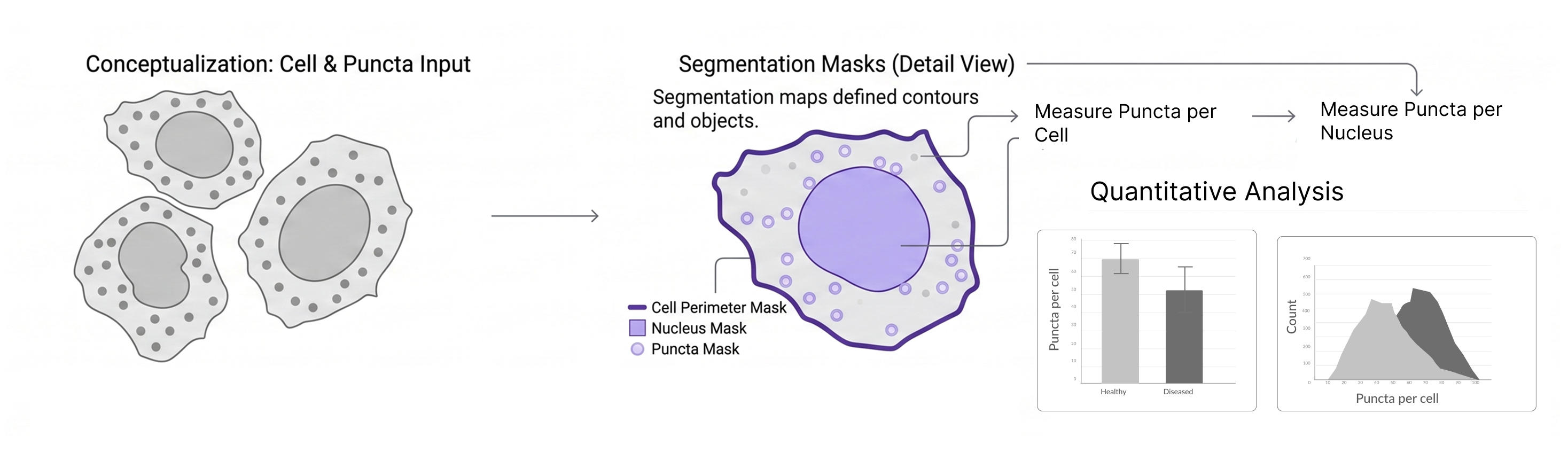
- Cell → puncta (puncta per cell)
- Tissue region → positive cell (positive cells per region)
- Tumor → immune cell (immune infiltration)
Size group ROI hierarchy — constrain where a child class is even segmented, so it only runs inside its parent region (e.g., cells only segmented within an organoid, positive cells only within a tissue region). This isn't just cleaner data — it saves analysis time and credits by skipping segmentation outside the region that matters.
Grow, shrink, and transform objects
Expand or contract detected objects by a set number of pixels to answer questions segmentation alone can't:
- Grow — associate cytoplasmic or extracellular signal with a nucleus, or connect nearby puncta to their nearest cell.
- Shrink — remove autofluorescence or edge artifacts near a boundary.
- Hollow or filled — turn a grown/shrunk region into a ring or a solid shape, then rename and recolor the resulting class.
See some examples
Quantify tissue hierarchy, automatically
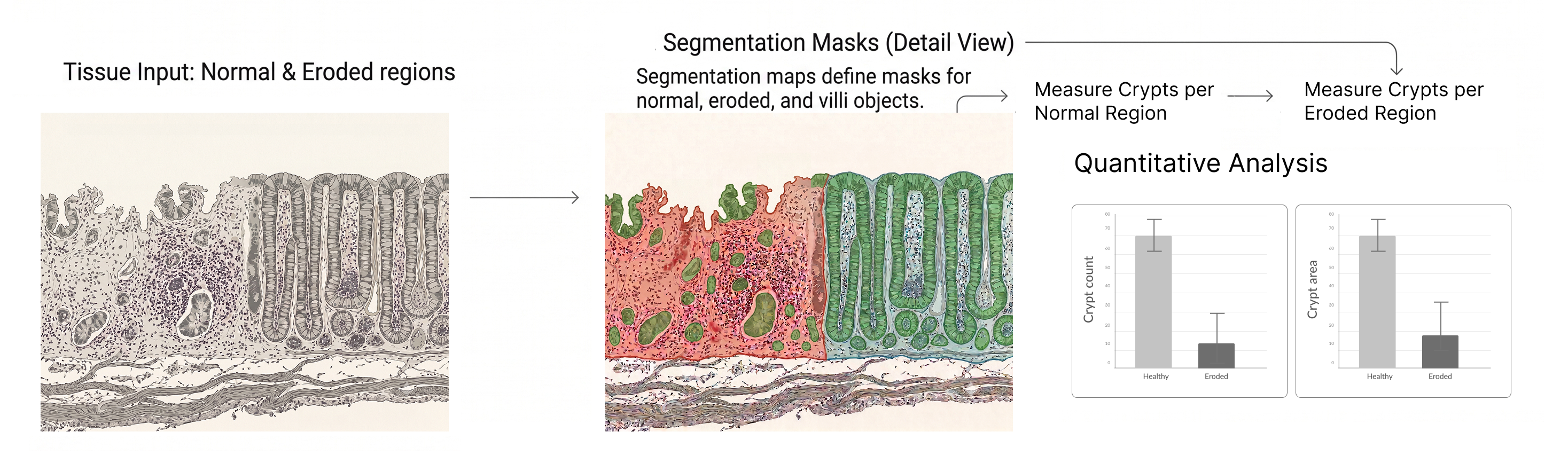
Segment normal and eroded tissue regions, then measure crypts, villi area, and nucleus count within each — automatically.
Turn your images into biological intelligence.
Start free, or talk to our team about your workflow.